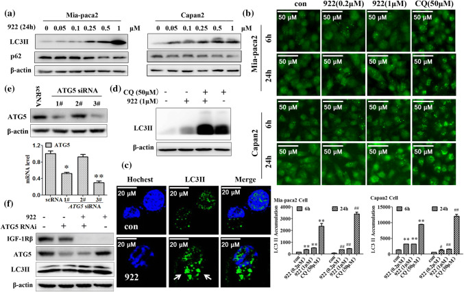
Fig. 4.
NVP-AUY922-induced IGF-1Rβ degradation is independent of macroautophagy. a Western blot analysis of the expression of the autophagy-related proteins LC3 II and p62 in Mia-paca2 and Capan-2 cells after treatment with the indicated doses of 922 for 24 h. β-actin served as a loading control. b Expression of LC3 II in Mia-paca2 and Capan-2 cells after treatment with 0.2 or 1 μM of 922 or 50 μM of CQ for 6 h or 24 h was assessed by immunofluorescence using a GE InCell Analyzer 1000. c Representative images of LC3 II puncta formation are presented after treatment with 1 μM of 922 for 24 h in Mia-paca2 cells (× 400). d Mia-paca2 cells were cultured in 1 μM of 922 for 24 h with or without CQ pretreatment (50 μM, 2 h). The LC3 II expression was detected using western blot analysis. e The mRNA level of ATG5 was silenced by siRNA in Mia-paca2 cells. f Knockdown of ATG5 by 50 nM targeted siRNA for 24 h in Mia-paca cells followed by treatment with 1 μM 922 for another 24 h. The expressions of ATG5 and IGF-1Rβ were detected by western blot analysis. The data are expressed as the mean ± SD (n = 3). **P ≤ 0.01 versus con of 6 h. #P ≤ 0.05, ##P ≤ 0.01 versus con of 24 h

