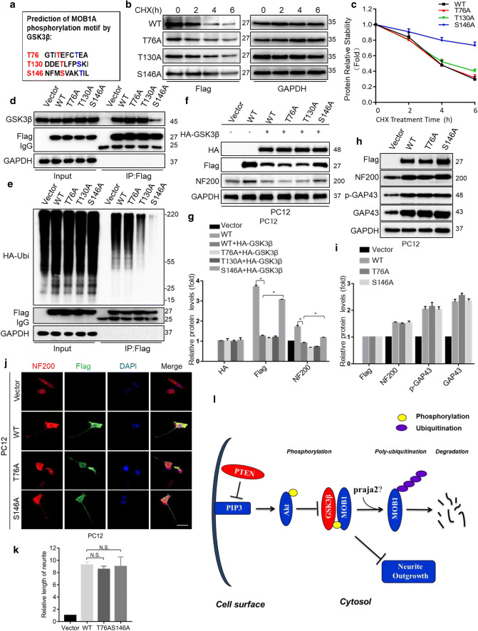
Fig. 7.
GSK3β phosphorylates MOB1 protein at the Ser146 site and promotes its proteolysis and working model. a Sequence analysis of mouse MOB1A identified three putative GSK3β target sites at T76, T130, and S146. NIH3T3 cells were transfected with Flag-tagged MOB1A (WT) and mutants (T76A, T130A or S146A), respectively. After 48 h, cells were treated with CHX (10 μM) for the indicated time points. Lysates were subjected to IB with Flag antibody and GAPDH served as an internal control (b). The curves of MOB1 relative stabilities were shown using the ratio of relative density normalized to that of 0-h group in three independent experiments (c). d Lysates from NIH3T3 cells expressing MOB1A (WT) or mutants T76A, T130A and S146A were subjected to IP using Flag antibody, followed by IB with GSK3β and Flag antibodies. Vector was used as a control. e MOB1A (WT) or mutants T76A, T130A and S146A were co-transfected with HA-Ubiquitin plasmids. Lysates from the above NIH3T3 cells were subjected to IP using Flag antibody, followed by IB with HA and Flag antibodies. Vector was used as a control. f, g HA-GSK3β and Flag-MOB1A or mutants were co-transfected into PC12 cells. Cell lysates were subjected to IB with anti-HA, anti-Flag, and anti-NF200. GAPDH served as an internal control (*P < 0.05 vs. WT group, ANOVA test followed by Dunnett’s post hoc test). h, i Lysates from PC12 cells expressing MOB1A (WT) or mutants T76A and S146A were subjected to IB with Flag, NF200, GAP43 and p-GAP43 antibodies. GAPDH served as an internal control. j, k Neurite outgrowth in PC12 cells was observed under different conditions. Representative immunofluorescence images of cells treated with anti-NF200 and anti-Flag for PC12 cells transfected with Flag-tagged MOB1A (WT) or mutants (T76A or S146A) are shown. Vector was used as a control. Scale bar, 100 μm (j). The quantification of the neurite length in PC12 cells stained with NF200-Cy3 (N.S. not significant vs. WT group, ANOVA test followed by Dunnett’s post hoc test) (k). l Working model. PTEN–GSK3β axis promotes ubiquitination and degradation of the MOB1 protein to suppress neurite outgrowth

