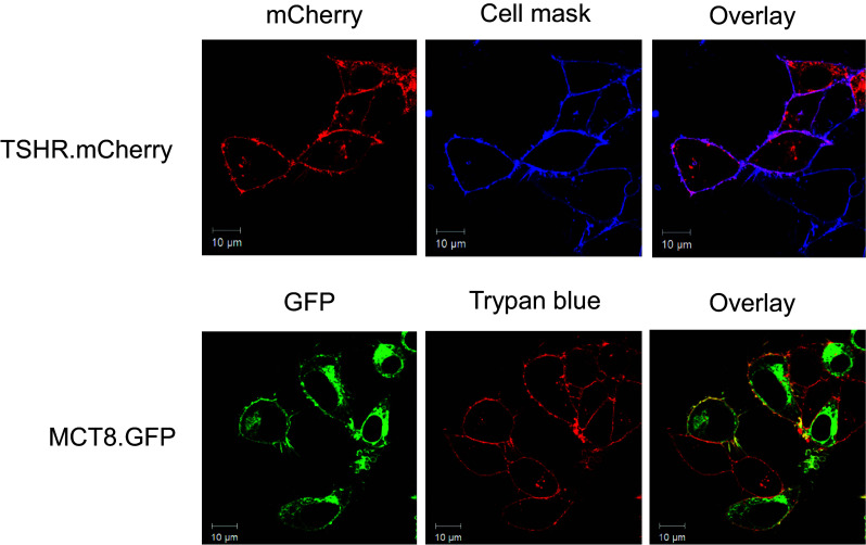
Fig. 2.

Localization of TSHR.mCherry and MCT8.GFP in the plasma membrane of transiently transfected HEK293 cells. The fluorescence signals of TSHR.mCherry (red, upper left panel) were co-localized with those of the plasma membrane stain CellMask Deep Red (blue, upper central panel). Purple color in the overlay (upper right panel) demonstrates that TSHR.mCherry is readily expressed in the plasma membrane. The fluorescence signals of MCT8.GFP (green, lower left panel) were co-localized with those of the plasma membrane stain trypan blue (red, lower central panel). Yellow color in the overlay (lower right panel) demonstrates that MCT8.GFP is expressed in the plasma membrane. Note that GFP and mCherry signals are only detectable in transfected cells whereas the dyes trypan blue and CellMask Deep Red stain each cell in the field of view. The scans show representative cells. Scale bar 10 μm. Similar data were obtained in four independent experiments
