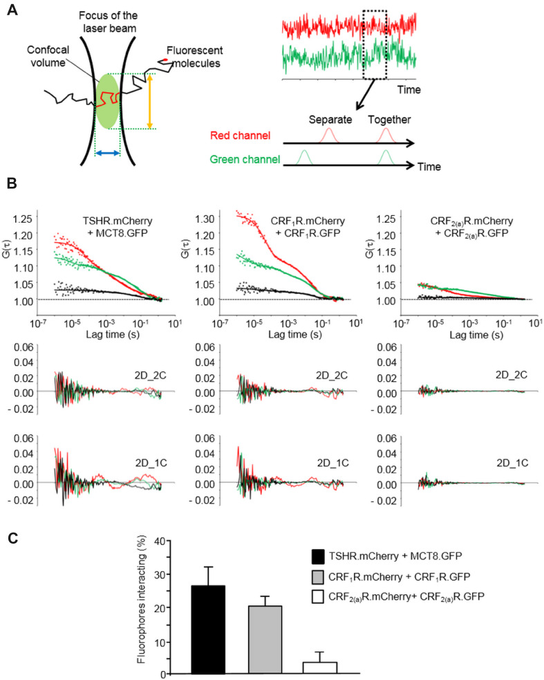
Fig. 3.
FCCS analysis of the TSHR.mCherry and MCT8.GFP interaction. a Principles of the FCCS methodology. A single fluorescent molecule diffusing through a very small confocal volume is recorded (left panel). The confocal volume is defined by the diameter of the microscopic lens (blue arrow) and axial structural parameter of the microscope objective (orange arrow). The focus of the laser beam is set to the plasma membrane. If two different fluorescent molecules were used, the fluctuations of molecules are recorded in two channels over time (right panel, red and green) and auto- and cross-correlation analyses can be performed to analyze whether the two fluorescent partners diffuse independently or together. b FCCS measurements using transiently co-transfected HEK293 cells expressing TSHR.mCherry and MCT8.GFP. The constructs CRF1R.mCherry/CRF1.GFP and CRF2(a)R.mCherry/CRF2(a)R.GFP were used as positive and negative controls respectively. The laser beam was focused at the plasma membrane. Representative auto- and cross-correlation data points and fitted curves for the analyzed constructs are shown. Auto-correlation data are depicted in red (mCherry) and green (GFP), cross-correlation data are shown in black. Curves were obtained by two-dimensional fits with two components (2D_2C) which gave the best results. Below each diagram, the residuals of this fitting are shown (auto-correlation data: GFP = green, mCherry = red). In addition, the residuals of suboptimal fits with 2 dimensions and 1 component (2D_1C) are depicted to demonstrate the necessity of 2D_2C fits. c Quantification of the TSHR.mCherry/MCT8.GFP interaction. The diagram shows the rescaled and distribution-corrected data of the FCCS experiments and indicates the portion of interacting TSHR.mCherry/MCT8.GFP fluorophores in the plasma membrane. In the case of CRF1R.mCherry/CRF1.GFP (positive control) and CRF2(a)R.mCherry/CRF2(a)R.GFP (negative control), the columns represent the portion of homodimers. Columns represent mean values ± SD of a total of 68 (TSHR.mCherry/MCT8.GFP), 51 (CRF1R.mCherry/CRF1.GFP) and 68 cells (CRF2(a)R.mCherry/CRF2(a)R.GFP). The total number of cells was collected in 5 independent experiments

