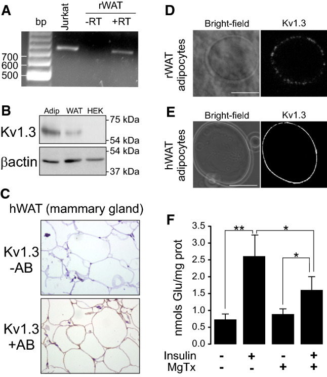
Fig. 4.

Human and rat WAT express Kv1.3 participating in insulin-activated glucose uptake in adipocytes. a RNA was extracted from rat WAT and RT-PCR was performed in the absence (−) or the presence (+) of retrotranscriptase (RT). Human Jurkat T-lymphocytes were used as a positive control. b Protein extracts from isolated rat adipocytes and WAT and human HEK 293 cells were analyzed for the expression of Kv1.3. β-actin was used as a loading and transfer control. c Human WAT from mammary gland biopsies was analyzed for the Kv1.3 staining in the absence (−) or the presence (+) of Kv1.3 antibody (AB). d Rat adipocytes from SVF were isolated and analyzed by immunohistochemistry for the presence of Kv1.3. e Adipocytes from subcutaneous tissue of human fat were stained for the expression of Kv1.3. Bars represent 50 µm. f Kv1.3 participates in insulin-dependent glucose uptake augmentation in adipocytes. Adipocytes were incubated with (+) or without (−) of 10 µM insulin for 30 min and glucose uptake was measured in the presence (+) or the absence (−) of 100 nM Margatoxin as described in the methods section. Values represent the mean ± SE, n = 4–6. *p < 0.05, **p < 0.01 Student’s t test
