Fig. 6.
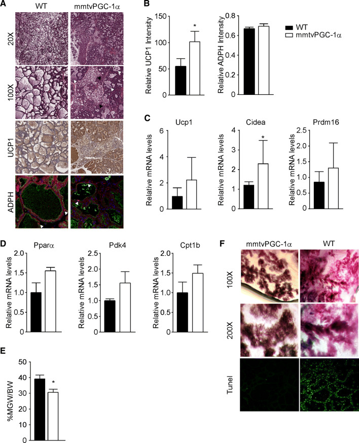
Stable overexpression of PGC-1α during lactation promotes apoptosis and mammary glands regression. a Mammary tissue sections from 10 days lactating WT and mmtvPGC-1α females mice stained with H&E (different magnification), with UCP1 immunohistochemistry, and ADPH immunofluorescence. In ×100 H&E, white arrows indicate the shedding of epithelial cells into the alveolar lumen; and black arrows represent brown adipocytes. Immunolocalization of ADPH was performed using Alexa 594-conjugated antibodies against the N-terminus of mouse ADPH (red, arrowed). Luminal borders of mammary alveoli were identified by staining with Alexa 488-conjugated WGA (green). Nuclei were stained with TO-PRO-3 (blue). b Quantification of UCP1 and ADPH immunostaining. Relative expression of c fat-browning related genes, Ucp1, Cidea and Prdm16, and d β-oxidation genes, Pparα, Pdk4 and Cpt1b, in lactating mammary glands evaluated by real time qPCR. TBP was used as housekeeping gene. e Inguinal mammary glands weight to body weight ratio (MGW/BW) of WT and mmtvPGC-1α lactating mice. f Low and high magnification of whole mount staining together with TUNEL staining of mammary glands isolated from lactating mmtvPGC-1α and WT mice. Comparison of wild-type and transgenic mice (n = 6, 7) was performed using Mann–Whitney U test. Data are expressed as mean ± SEM (*p < 0.05; **p < 0.01)

