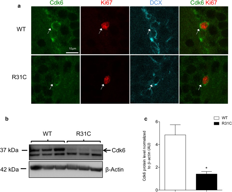
Fig. 3.
Cdk6R31C mutation decreases Cdk6 protein level in hippocampal progenitors. a Representative single-plane confocal images of Cdk6 (green)/Ki67 (red)/DCX (cyan) triple staining in the SGZ of WT and R31C mice. White arrows point to triple-labeled cells. b Western blot analysis of Cdk6 expression in protein extracts from P6 to P7 WT and R31C hippocampi. Each well corresponds to one independent animal. β-actin serves as a loading control. The black arrow points to the specific Cdk6 band. c Histogram showing the quantification of the western blot depicted in b. Data are presented as mean ± SEM and were analyzed by an unpaired two-tailed Student’s t test. N = 3 mice per genotype. *p < 0.05. AU arbitrary unit

