
Fig. 1.
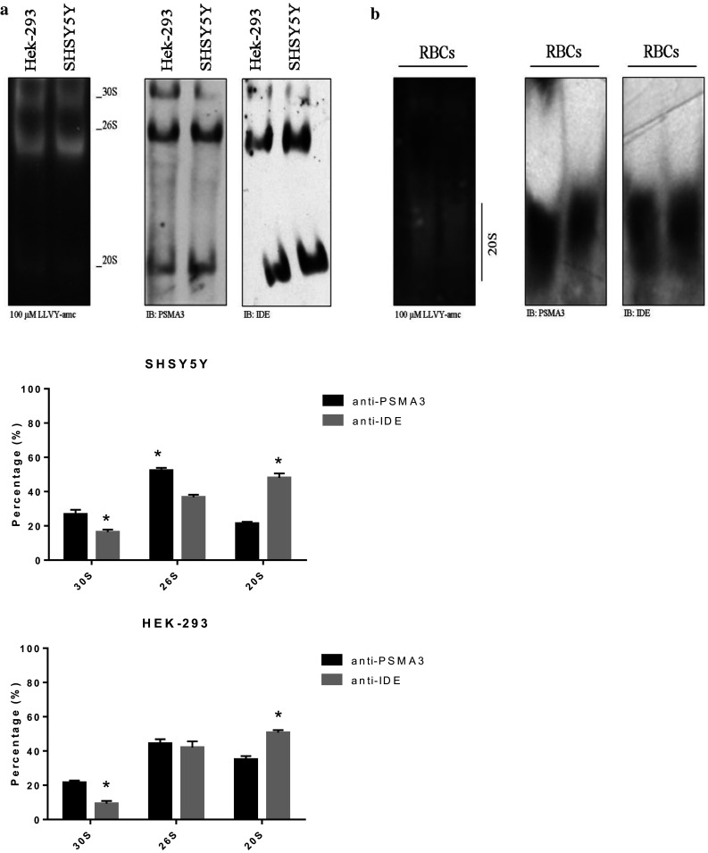
The extracts of the SHSY5Y and HEK-293 cells (left panel) and of the RBCs (right panel) were resolved by native gel electrophoresis and the proteasome particles probed with the LLVY-amc fluorogenic substrate. The proteasome particles were probed with an anti-IDE and anti-PSMA3 antibody by WB. The relative abundance of the three particles was determined through a densitometric analysis of the WB bands (bottom panel). A representative immunoblot is shown. Values reported are the mean ± SE of five independent experiments. *Significantly different from the PSMA3 staining (p < 0.001, one-way ANOVA, followed by Tukey’s test, n = 15)