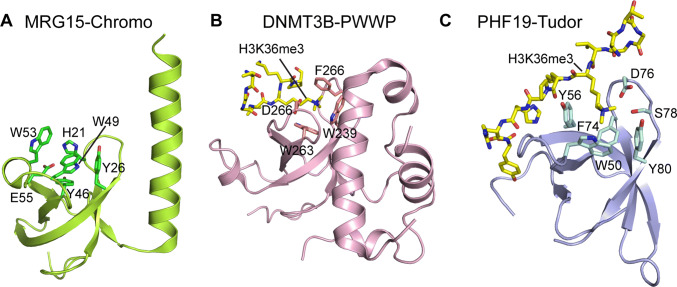
Fig. 1.
Structures of the H3K36 methylation-specific ‘reader’ domains, as exemplified by a the chromodomain of MRG15 (PDB: 2F5K), b the PWWP domain of DNMT3B (PDB: 5CIU) and c the Tudor domain of PHF19 (PDB: 4BD3). The H3K36me3-engaging residues are labeled in each panel, with the histone peptide shown in gold

