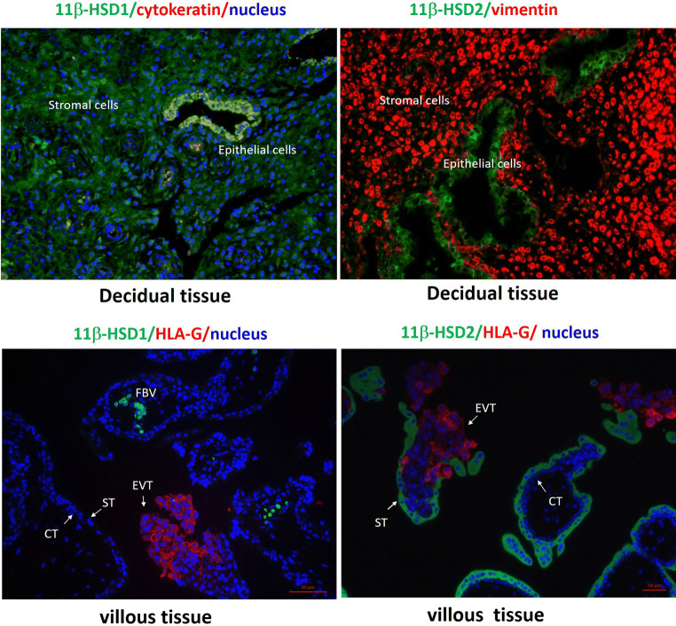
Fig. 1.
Distribution of 11β-HSD1 and 2 in human decidua and chorionic villous tissues during early gestation. Top panel: Decidual epithelial cells are cytokeratin 7 positive and stromal cells are vimentin positive. Bottom panel: Villous trophoblasts are HLA-G negative while extravillous trophoblasts are HLA-G positive. CT cytotrophoblast; ST syncytiotrophoblast; EVT extravillous trophoblast; FBV fetal blood vessels. Based on the work presented in the Ref. [46]. Human tissue collection was approved by the Ethics Committee of Ren Ji Hospital, Shanghai Jiao Tong University School of Medicine with informed consent

