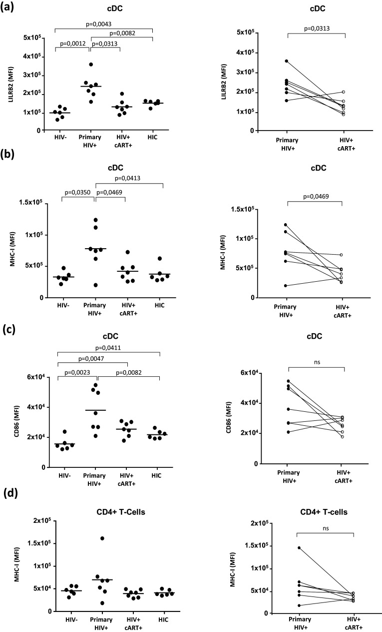
Fig. 1. Characterization of LILRB2 and MHC-I expression level on cDCs from patients during the early phase of HIV-1 infection.
a–c Analysis of LILRB2, MHC-I, and CD86 surface expression on cDCs from blood samples of early HIV-1 infected patients before (primary HIV+) or after 1 year of cART (HIV+ cART+), HIV-1 infected elite controller patients (HIC), and HIV-1 non-infected controls (HIV-). d Analysis of CD4+ T cells in these samples to evaluate surface expression of MHC-I. Data are represented as mean fluorescence intensity with statistical analysis performed using the Mann–Whitney U test between HIV- (n = 6), HIC (n = 6), and primary HIV+ patients (n = 7) and the Wilcoxon signed-rank test for HIV+ primary patients before and after cART (p < 0.05 is considered significant)

