
Fig. 2. Comparison of the LILRB2 expression pattern between human and cynomolgus macaque immune cell subsets.
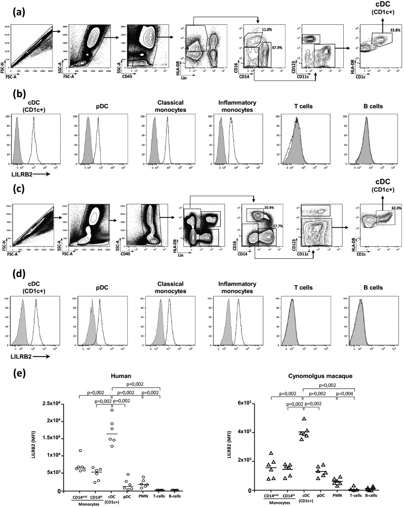
Flow cytometry gating strategy to characterize blood immune cell subsets in humans (a) and cynomolgus macaques (c). Surface expression of LILRB2 was assessed using monoclonal antibodies against human (clone 42D1) or cynomolgus macaque (clone 17E5.3E9) LILRB2. The distribution of LILRB2 expression is shown for various immune cell subsets, including cDCs, pDCs, classical (CD14high CD16−) and inflammatory (CD14mid CD16high) monocytes, T and B cells in healthy (b) human donors and (d) cynomolgus macaques. e Comparison of LILRB2 surface expression on whole blood immune cell subsets of healthy human donors (n = 6) and cynomolgus macaques (n = 6). Results shown are representative of at least three independent experiments. Statistical analyses were carried-out using the Mann–Whitney U test for both human and cynomolgus macaque samples (p < 0.05 is considered significant)