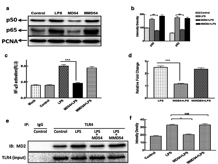
Fig. 6.
Study of effect of MD54 and MMD54 on the translocation of NF-κB into the nucleus in LPS-stimulated THP-1 and HEK-293T cells and MD54-induced attenuation of LPS-induced interaction between MD2 and TLR4. a Western blot of the nuclear fraction of LPS-stimulated THP-1 cells for p65/p50 subunit of NF-κB in the absence and the presence of MD54 and MMD54. b Intensity graph. **p < 0.01, one-way analysis of variance using Turkey’s test was performed using Graph Pad Prism Version 5.00 vs. LPS, c NF-κB activation in HEK-293T transfected cells. d Relative fold change in NF-κB activation in HEK-293T after treatment of LPS and peptides (RLU control = 1). Treatment of the peptides is as marked in the figure, whereas control and LPS represent untreated and LPS-treated THP-1 and HEK-293T without the addition of peptides (Mock represent un-transfected cell treated with peptides). ***p < 0.001, Student t test was performed using Graph Pad Prism Version 5.00 vs. LPS. e Immunoprecipitation assay showed that MD54 pretreatment (10 μM) significantly reduced the formation of the TLR4–MD2 complex induced by 100 ng ml−1 LPS. f Densitometric analysis was done using the ImageJ software. Data represented are WB analyses of at least two independent experiments (*p < 0.05)

