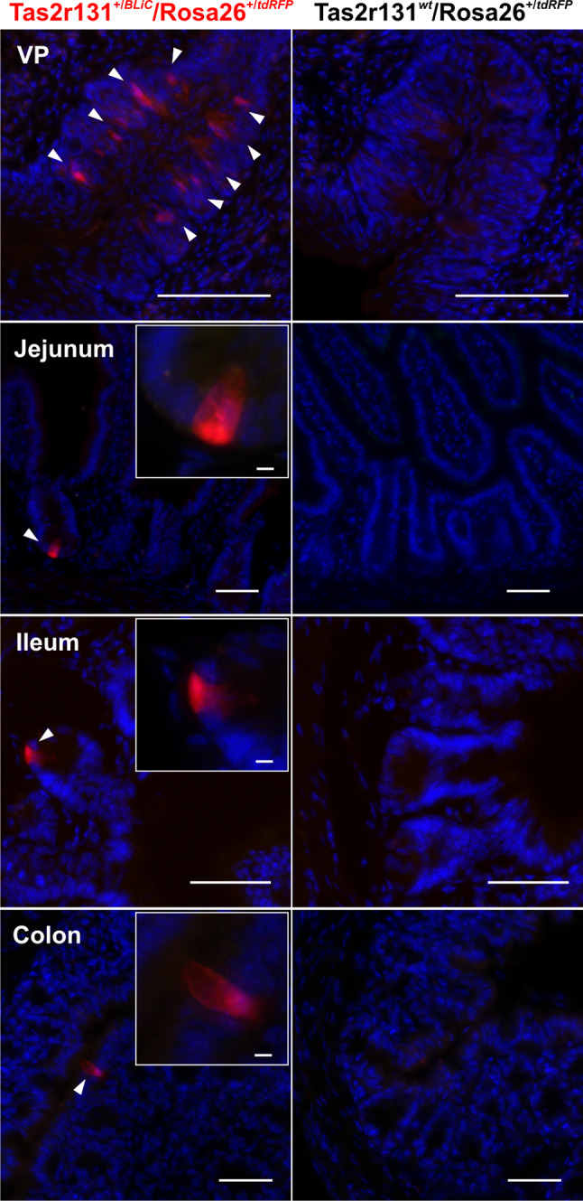
Fig. 1.

Histochemical characterization of GI tissues from Tas2r131+/BLiC/Rosa26+/tdRFP and Tas2r131wt/Rosa26+/tdRFP control animals. Cryostat sections of 14 μm thickness were stained with DAPI and subjected to fluorescent scans with a MIRAX Midi system. Arrowheads point to Tas2r131+ cells in red (Cy3 filter), DAPI-stained nuclei are depicted in blue (DAPI filter). Green (FITC filter) was used as background. The vallate papillae (VP) served as a positive control for the expression of the Tas2r131 gene in taste cells. Tas2r131+ cells are found in the jejunum, ileum, and colon as already reported (Prandi et al., 2013). Scale bars 50 µm. Inserts, scale bar 5 µm, show a magnification of the Tas2r131+ cells
