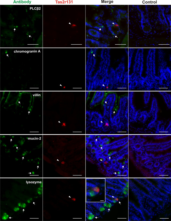
Fig. 2.
Immunohistochemical identification of the Tas2r131+ cells with intestinal-specific cell and taste markers in the ileum of Tas2r131+/BliC/Rosa26+/tdRFP animals. Representative microphotographs showing ileal Tas2r131+ cells appearing in the mucosal layer of the intestinal wall, and the main mucosal cell types as identified by specific antibodies. Cryostat sections of 14 μm thickness were stained with the corresponding antibodies (and corresponding controls) for known markers and then subjected to fluorescent scan with a MIRAX Midi system. Arrowheads point to Tas2r131+ cells in red (Cy3 filter), arrows to antibody-labelled cells in green (FITC filter), DAPI-stained nuclei are depicted in blue (DAPI filter). The insert shows a magnification of a Tas2r131+ cell positive for lysozyme labelling. Control reactions for each antibody (blocking peptide for villin and lysozyme, minus antibody control for chromogranin A, mucin-2, and PLCβ2) are shown on the right column. Scale bars 50 µm, 10 µm (inserts)

