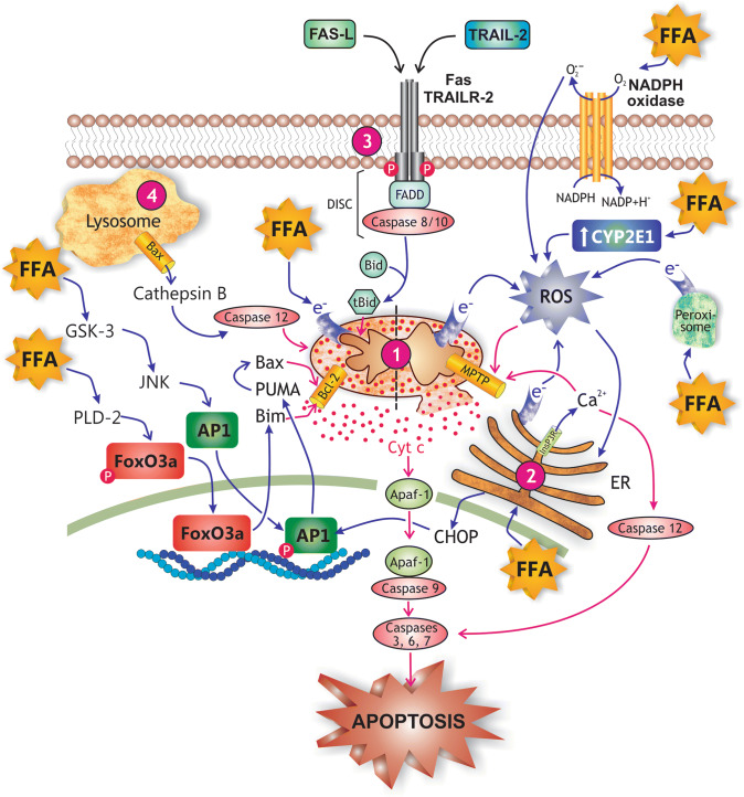
Fig. 7.
Main mechanisms of free fatty acid (FFA)-induced hepatocellular apoptosis in NASH. FFAs promote apoptosis by virtually all known apoptotic pathways. (1) The intrinsic or mitochondrial pathway of apoptosis, which is activated by FFA-induced cellular stress, and mediated by the release of cytochrome c (Cyt c) from the mitochondrial intermembraneous space to cytosol; Cyt c promotes binding of pro-caspase 9 to apoptosis protease-activating factor-1 (APAF-1), pro-caspase 9 activation by autocatalysis, and finally, recruitment and activation by caspase 9 of the executioner caspases 3, 6, and 7. FFAs activate this pro-apoptotic pathway by a activating the glycogen synthase kinase-3 (GSK-3)/JNK and the protein phosphatase 2A (PP2A) pathways, which causes the transcription factors FoxOa3 and AP-1 to translocate to the nucleus and to induce transcription of genes encoding pro-apoptotic proteins of the Bcl-2 family (PUMA, Bim); Bim is a pore-forming protein whereas PUMA activates the pore-forming protein Bax by binding inhibitory members of the Bcl-2 family, which release Bax from this inhibitory constraint (see mitochondrion, left side), and b by promoting the formation of mitochondrial permeability transition pores (MPTP), a process facilitated by reactive oxygen species (ROS) generated from leakage of electrons from the respiratory chain in mitochondria, from oxidases in peroxisomes and endoplasmic reticulum (ER) involved in FFA β- and ω-oxidations, and from induction of pro-oxidizing enzymes, such as NADPH oxidase 4 (NOX4) and CYP2E1 (see mitochondrion, right side); mitochondria permeabilization also exacerbates the leakage of electrons from the mitochondrial electron transport chain and further cytosolic ROS generation. (2) The endoplasmic reticulum (ER) stress pathway of apoptosis, which is induced by both FFAs and ROS, and mediated by a inositol 1,4,5-triphosphate receptor (Ins3PR)-induced Ca2+ release to cytosol and further Ca2+-dependent activation of the executor caspase 12, b Ca2+-dependent MPTP formation in mitochondria, and c ER stress-induced CCAAT-enhancer-binding protein homologous protein (CHOP) expression, which enhances AP-1 transcriptional activation. (3) The extrinsic pathway of apoptosis, which is induced sequentially by a binding of pro-inflammatory cytokines (e.g., Fas-L and TRAIL) to their respective plasma membrane receptors (Fas and TRAILR), b autophosphorylation of these receptors, c association of the activated receptors with FADD and with pro-caspases 8 and 10 to form the death complex DISC, d proteolytic activation of these pro-caspases, and e caspase-dependent cleavage of Bid to truncated Bid (tBid), which promotes Bcl-2-dependent mitochondrial pore formation. (4) The lysosomal pathway of apoptosis, which involves FFA-induced Bax translocation to lysosomes and further cytosolic release of cathepsin B; this protease activates caspases 12, which would interact directly with mitochondria to induce membrane permeabilization

