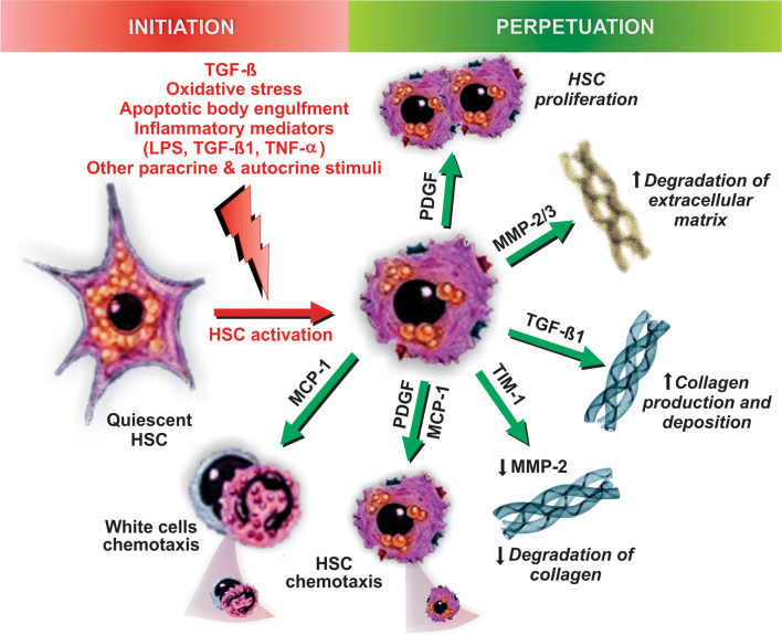
Fig. 9.
Pathways of initiation and perpetuation of fibrosis mediated by hepatic stellate cell (HSC) activation. Following liver injury, a number of hormonal, paracrine, and autocrine stimuli induce HSC activation, a process initiated by the transition from quiescent, vitamin A-rich HSCs into fibrogenic, proliferative ones; TGF-β produced by Kupffer cells is the main stimulus that drives this phenotypic change. This ‘initiation phase’ is followed by the ‘perpetuation phase’, in which this phenotype is preserved and potentiated by additional stimuli that promote HSC proliferation (via the platelet-derived growth factor [PDGF]), sustained collagen production and deposition (via TGF-β), matrix degradation (via matrix metalloproteinases 1 and 3 [MMP-1/3]), inhibition of collagen degradation (via the MMP-2 inhibitor, tissue inhibitor of metalloproteinase-1 [TIM-1]), HSC chemotaxis (via PDGF and monocyte chemoattractant protein-1 [MCP-1]), and white cell chemoattraction (via MCP-1)

