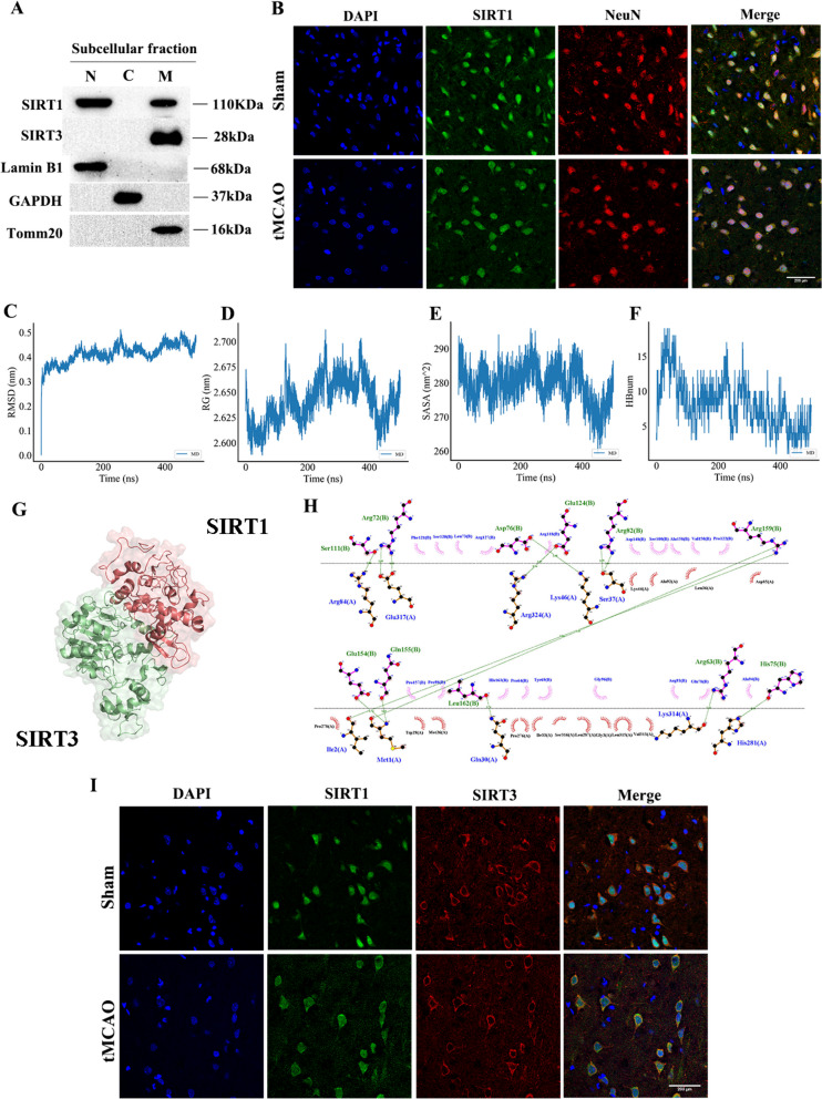
Fig. 1.
The interaction between SIRT1 and SIRT3 before and after CI/R. A Representative immunoblots of SIRT1 and SIRT3 in each subcellular fractionation in neurons. n = 3. B Representative confocal images of co-localization of SIRT1 and NeuN (neuron marker) (63 × objective). n = 3. Scale bar: 200 μm. C RMSD. D Rg. E SASA. F HBnum. G SIRT1 bound to SIRT3 in a 3D space. H 2D ligand-residue analysis. I Representative confocal images of co-localization of SIRT1 and SIRT3 (63 × objective). n = 3. Scale bar: 200 μm

