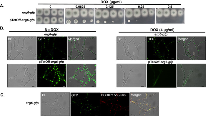
Fig. 6. Erg6 localizes to lipid droplets in A. fumigatus hyphae.
A Spot-dilution cultures, performed as described in Fig. 1B, indicate that fusion of egfp to the 3’ end of erg6 in either the parent or pTetOff-erg6 background does not negatively affect erg6 function. Note similarities to growth in untagged strains (Fig. 1B). B Mature mycelia were developed in GMM broth using the indicated doxycycline (DOX) concentrations for 16 h at 37 °C. Fluorescent images were captured using GFP filter settings. C Co-localization of Erg6-GFP to A. fumigatus lipid droplets using the droplet marker, BODIPY 558/568. Conidia of the erg6-gfp strain were cultured to mature hyphal development and subsequently stained with 1 µg/ml BODIPY 558/568 C12 for 20 min at room temperature. Images were captured using GFP and TRITC filter settings, respectively. Scale bar = 10 µm. Microscopy experiments were completed three times independently with similar results.

