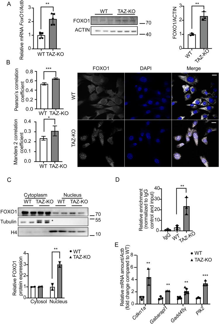
Figure 3.
Upregulation of FOXO1 activates PDK4 transcription in TAZ-KO cells. (A) Total mRNA and whole-cell lysate were extracted from both WT and TAZ-KO myoblasts, and the mRNA and protein levels of FOXO1 were measured using real-time qPCR and WB analysis, respectively. Data are presented as fold-change relative to WT, and each lane represents an individual biological replicate. (B) Right: The subcellular localization of FOXO1 was detected through immunofluorescence (IF) confocal microscopy, with representative figures showing the relative distribution of FOXO1 in WT and TAZ-KO myoblasts. Left: Quantification of colocalization between FOXO1 and DAPI based on Pearson's and Mander's 2 colocalization analysis56,57. The scale bar equals 20 µm. (C) Nuclear protein fractionation was performed, followed by WB analysis using tubulin and histone 4 (H4) as internal controls for cytoplasmic and nuclear fractions, respectively. * indicates PDK4 band residue after stripping the membrane. (D) Chromatin immunoprecipitation/qPCR was performed in myoblasts with either an IgG control or anti-FOXO1 antibody. The Pdk4 promoter was detected using the specific primers listed in “Experimental procedures”. (E) Real-time qPCR was used to measure mRNA of the FOXO1 targets Cdkn11 (Cyclin Dependent Kinase Inhibitor 1A), Gabarapl1 (Gamma-aminobutyric acid receptor-associated protein-like 1), Gadd45γ (Growth Arrest and DNA Damage Inducible γ), and Plk2 (Polo Like Kinase 2), using Actb as the internal control. Data points represent mean ± S.D. (error bars) for each individual biological replicate of each group. * 0.05 < p < 0.1, ** 0.01 < p < 0.05, *** 0.001 < p < 0.01.

