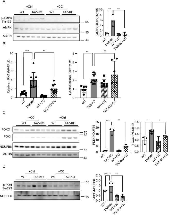
Figure 5.
AMPK-mediated upregulation of FOXO1 increases PDK4 in TAZ-KO cells. (A) Phosphorylation of AMPK residue Thr172 (p-AMPK) was assayed in WT and TAZ-KO myoblasts treated with the AMPK inhibitor compound C (CC) or vehicle. 10 μg of total protein was loaded for each sample, and ACTIN was used as an internal control. Representative images and quantitative analysis are shown. (B) Total mRNA was extracted from myoblasts, and Pdk4 and Foxo1 mRNA levels were measured via real-time qPCR following treatment with CC (10 μM, 16 h) or vehicle. (C) Protein expression of FOXO1 and PDK4 in myoblasts was measured via WB analysis following treatment with CC (10 μM, 16 h) or vehicle. Representative images and corresponding quantitative analyses are shown. (D) In myoblasts, p-PDH was measured by WB analysis following treatment with CC (10 μM, 16 h) or vehicle. Representative images and corresponding quantitative analyses are shown. Data points represent mean ± S.D. (error bars) for each individual biological replicate of each group. * 0.05 < p < 0.1, ** 0.01 < p < 0.05, *** 0.001 < p < 0.01.

