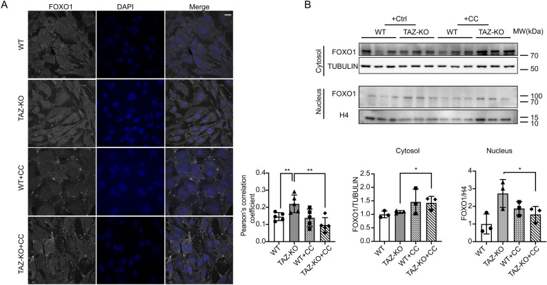
Figure 6.
AMPK activation promotes FOXO1 nuclear translocation in TAZ-KO cells. (A) IF staining was used to show the localization of FOXO1 in myoblasts following treatment with CC (5 μM, 16 h) or vehicle. Scale bar = 10 μm. Pearson's colocalization analysis between DAPI and FOXO1 (bottom); n = 5. (B) FOXO1 protein levels were assayed in nuclear and cytoplasmic cellular fractions following treatment with CC (5 μM, 16 h) or vehicle. Tubulin and histone H3 were used as internal controls for cytoplasmic and nuclear fractions, respectively. Corresponding quantitative analyses are also presented (bottom right). Data points represent mean ± S.D. (error bars) for each individual biological replicate of each group. * 0.05 < p < 0.1, ** 0.01 < p < 0.05, *** 0.001 < p < 0.01.

