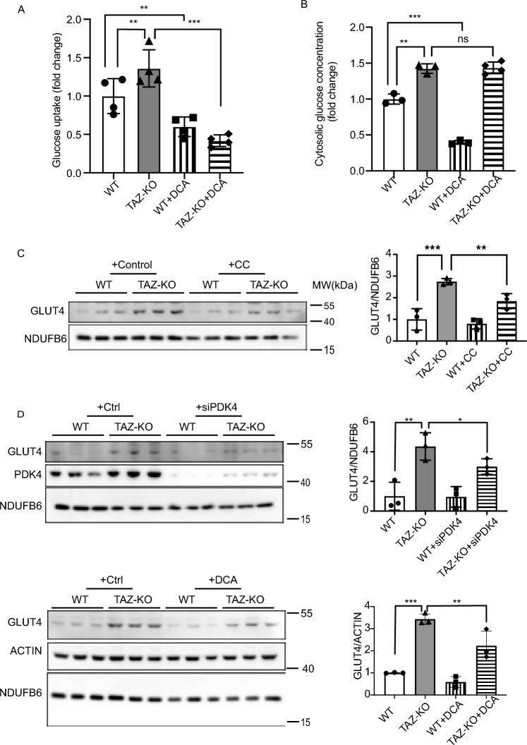
Figure 7.
Glucose utilization is dysregulated in TAZ-KO cells. (A) Myoblasts were incubated with 1 mM 2DG. The glucose uptake rate was determined by measuring the amount of 2DG taken up by the cells, with values normalized to total protein in each sample before analysis. (B) Cytosolic glucose concentration was measured according to “Experimental procedures”. GLUT4 protein expression was measured by WB in myoblasts treated with either 10 μM CC (C) or 5 mM DCA for 16 h or PDK4-targeted siRNA for 24 h (D). NDUFB6 and ACTIN were used as internal controls. The data points represent means ± S.D. (error bars) for each individual biological replicate of each group. * 0.05 < p < 0.1, ** 0.01 < p < 0.05, *** 0.001 < p < 0.01.

