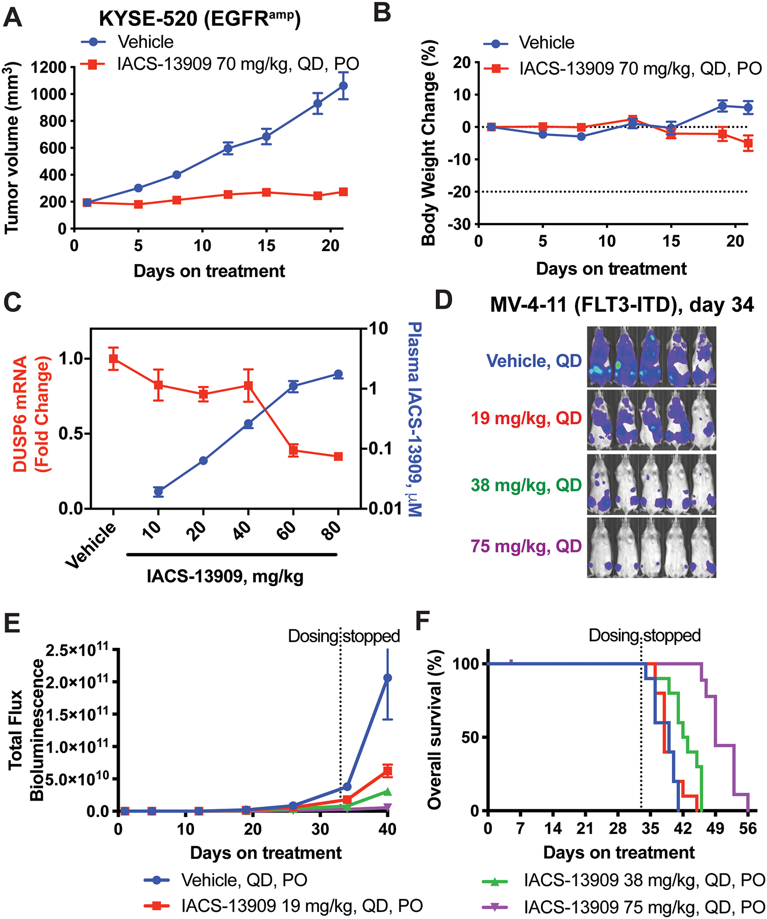
Figure 2. IACS-13909 suppresses proliferation and MAPK pathway signaling of RTK-activated tumors in vivo.

Tumor growth curve (A) and mouse body weight change (B) of the KYSE-520 subcutaneous xenograft model in mice, when treated with either vehicle (0.5% methylcellulose) or IACS-13909 at 70 mg/kg QD orally for 21 days. N=9 mice per group. (C) Plasma concentration of IACS-13909 (blue curve) and DUSP6 mRNA level in KYSE-520 subcutaneous tumor samples (red curve) from mice treated with vehicle or IACS-13909. Plasma and tumor samples were harvested 24 hours after a single dose treatment. N=3 mice/group/timepoint. (D-F) Anti-tumor efficacy of IACS-13909 on MV-4–11 orthotopic mouse model. Mice were injected with MV-4–11-Luc cells through tail vein, and treated with different doses of IACS-13909 QD orally. N=10 mice/group. (D) Representative mouse images from bioluminescence imaging indicating tumor volume on day 34. (E) Quantitated tumor volume determined by bioluminescence imaging. (F) Kaplan-Meier curve showing the overall survival of the mice with or without IACS-13909 treatment. The dotted vertical line indicates when dosing stopped.
