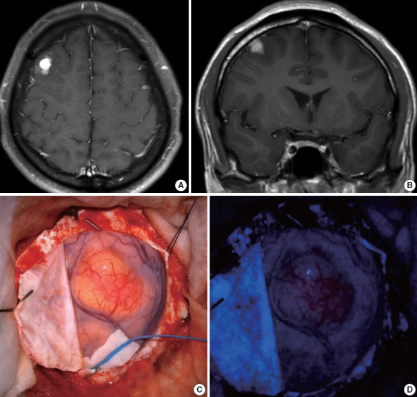
Fig. 1.
Contrast-enhanced magnetic resonance imaging (MRI) glioma study (3.0T) and intraoperative surgical field view. T1 axial (A) and coronal (B) preoperative MRI of the 1.5 cm vividly enhancing mass in the right frontal lobe with no intralesional hemorrhage or calcification. (C) Intraoperative surgical field view during the right frontal craniotomy and tumor removal. (D) Intraoperative 5-aminolevulinic acid uptake in the tumor.

