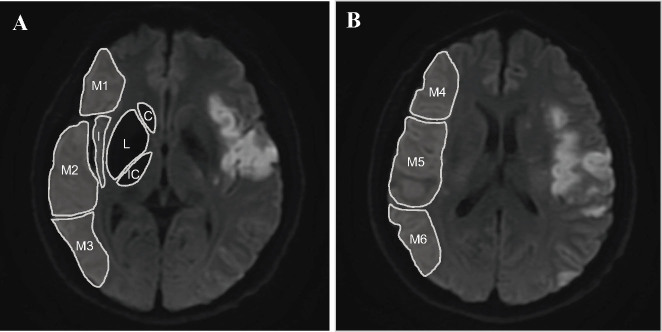
Figure 1.
Illustration of the assessment of DWI-ASPECTS. A shows the ganglionic level, which consists of 3 deep subregions (C: caudate nucleus, L: lenticular nucleus, and IC: internal capsule) and 4 cortical subregions (I: insula, M1: anterior inferior frontal lobe, M2: temporal lobe, and M3: inferior parietal and posterior temporal lobe). B shows the supraganglionic level, including 3 cortical subregions superior to M1, M2, and M3 (M4: superior anterior frontal lobe, M5: precentral and superior frontal lobe, and M6: superior parietal lobe). The DWI-ASPECTS score of this patient was rated as 5. DWI-ASPECTS: Alberta Stroke Program Early CT Score on MR-diffusion weighted imaging.

