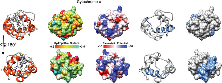
Fig. 2.
Cytochrome c and the derived CYCS77–101. Tridimensional surfaces of cytochrome c (CYCS UniProt structure identifier 3ZCF [98]); lower line of molecules corresponds to opposite face of the molecule depicted on first line; in blue, in rightmost two structures, segment of residues 77–101, which has bioportide properties (cell penetration and cytotoxicity). Electrostatic surface colored according to Coulombic electrostatic potential, e = 4r, thresholds ± 5 kcal mol−1 e−1 at 298 K. Hydrophobicity surface colored following the Hessa and von Heijne hydropathic scale thresholds (dark orange most hydrophobic; white 0; aquamarine most hydrophilic) as described by Hessa et al. [99]. Molecular graphics and analyses performed using the UCSF Chimera package software [100]

