Fig. 3.
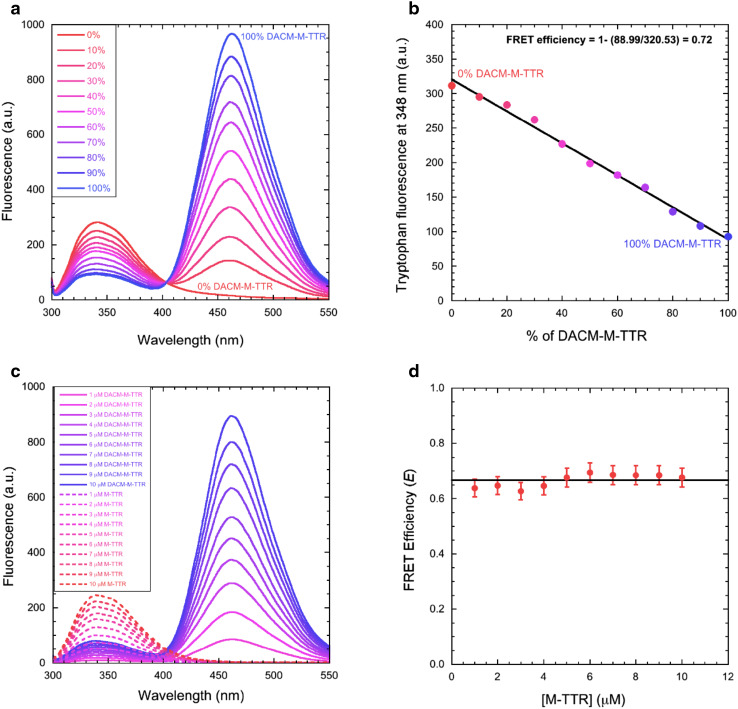
FRET of native M-TTR. a Fluorescence spectra of mixtures of M-TTR and DACM-M-TTR at the indicated percentages of the latter, at 3 µM total protein concentration, pH 7.4, 25 °C. b Tryptophan fluorescence emission at 348 nm versus the percentage of DACM-M-TTR. The straight line represents the best fit of the data points to a linear function. The equation indicates how E was determined. c Fluorescence spectra of M-TTR (dashed lines) and DACM-M-TTR (continuous lines) at various protein concentrations ranging from 1 to 10 µM, pH 7.4, 25 °C (excitation 290 nm). d Plot of E versus M-TTR concentration. The straight line represents the average value

