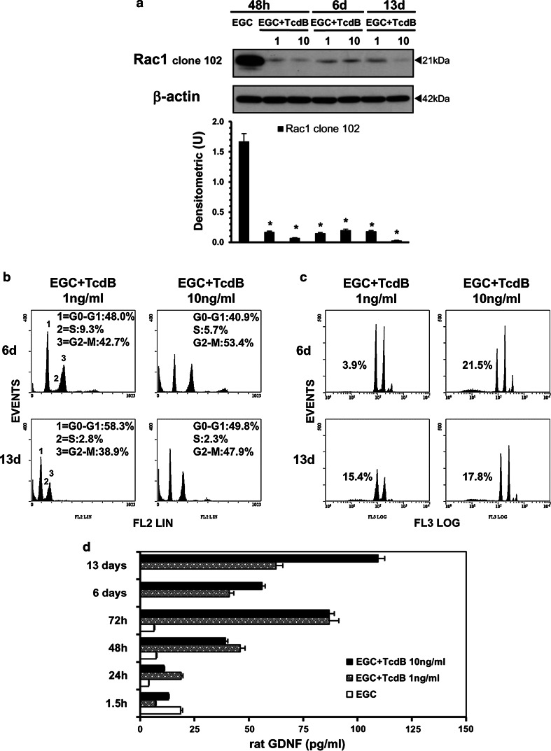
Fig. 16.
TcdB causes persistent alterations in EGCs. Control EGCs or EGCs treated with TcdB (1, 10 ng/ml) at 48 h were washed to remove TcdB and were then incubated for 6 or 13 days. The cells or supernatants were recovered, a at 48 h, 6 or 13 days for the preparation of cell lysates and Western blot analysis of Rac1 glucosylation. The graph represents the densitometric analysis of Rac1 relative to β-actin. *P < 0.01 TcdB-treated EGCs at different times versus control EGCs at 48 h; b at 6 or 13 days to evaluate the cell percentages in cell-cycle phases by flow cytometry (the results of one experiment, representative of four for each time, are shown); c at 6 days or 13 days to evaluate the percentage of hypodiploid nuclei via flow cytometry (the results of one experiment, representative of four for each time, are shown); d at 1.5–72 h, 6 or 13 days to evaluate GDNF secretion in culture supernatants by ELISA. The data are the mean ± standard deviation of three experiments performed in triplicate

