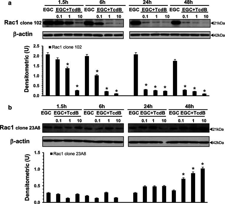
Fig. 2.
TcdB induces Rac1 glucosylation in EGCs. Lysates from control EGCs and EGCs treated with TcdB (0.1–10 ng/ml) prepared at 1.5–48 h were subjected to SDS-PAGE. The filters were: a probed with anti-Rac1 clone 102, then stripped and probed with anti-β-actin; b cut to around 30 kDa and the bottom sections were probed with anti-Rac1 clone 23A8 and top sections were probed with anti-β-actin. The graphs represent the densitometric analysis of each protein relative to β-actin. *P < 0.01 TcdB-treated EGCs versus control EGCs

