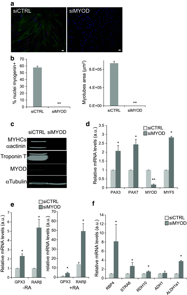
Fig. 6.
MYOD silencing blocks human myoblast differentiation and activates the retinoic-acid-signalling pathway. a Representative images (six fields from each condition) showing immunofluorescence analysis of the expression of myogenin (red) and Troponin T (green) in human myoblasts transfected with siCTRL (control) or siMYOD. DNA was stained with DAPI (blue). b Quantification of the percentage of myogenin-positive nuclei relative to all nuclei (left panel) and of the average area of myotubes in the cultures shown in a. c Western blot analysis of the expression of MyoD and the muscle differentiation markers MYHC slow (MYHCs), Troponin T, and alpha-actinin (α-actinin) in human myoblasts transfected with siCTRL or siMYOD. Alpha tubulin (α-tubulin) was used as loading control. d RT-qPCR analysis of the expression of myogenic determination genes in human myoblasts transfected with siMYOD relative to myoblasts transfected with sictrl (set to 1). e RT-qPCR evaluation of the expression of the RA-target genes RARβ and GPX3 in human myoblasts transfected with siCTRL or siMYOD incubated (+RA) or not (−RA, 0.1% DMSO) with RA. f RT-qPCR analysis of the expression of genes involved in the RA-signalling pathway in myoblasts transfected with siMYOD relative to control myoblasts (siCTRL). p ≤ 0.05 (*), p < 0.01 (**). Scale bars 10 μM

