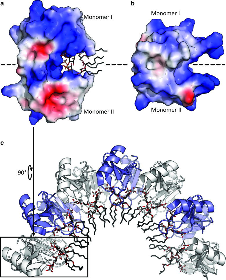
Fig. 6.
Defensin dimerisation and lipid-mediated oligomerisation. a The plant C8 defensin NaD1 (PDB:4CQK) forms a homodimer that binds negatively charged phospholipid head groups via a cationic grip [94]. b The human β-defensin HBD-2 (PDB:1FD4) forms a structurally similar dimer [102]. Protein surface charge is indicated by blue (positive) and red (negative). Lipids are shown as sticks with phosphate in white and oxygen in red. c NaD1 dimers assemble into an arching oligomeric structure after interaction with the anionic head groups of PIP2 within an extended cationic groove on the surface of the NaD1 oligomer (PDB:4CQK). Alternating dimers in white and blue

