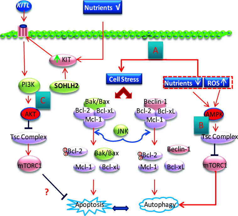
Fig. 4.
Schematic representation of autophagy and apoptosis regulation as well as the meiosis initiation mechanism during CBD and PF formation. a Cellular stress induced by starvation, ROS (reactive oxygen species), and other factors causes the displacement of Bcl-2 from either Beclin-1 or Bax, thereby triggering autophagy or apoptosis, respectively. Activated JNK is responsible for the phosphorylation of Bcl-2. The autophagy–apoptosis interactions are not fully understood. b Amino acid deprivation, hypoxia, and an elevated AMP/ATP ratio activate AMPK (5′-AMP-activated protein kinase), which in turn activate TSC2 (tuberous sclerosis complex 2), thus preventing the action of mTORC1 from inhibiting autophagy. c Increase of KIT levels in oocytes induced by KITL or SOHLH2 may activate mTORC1 to inhibit apoptosis through the PI3K/AKT signaling pathway

