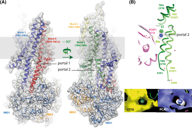
Fig. 4.
Lateral portals. a 3D structure model (ribbon and surface representation) of the possible open form of the CFTR MSD:NBD assembly, also shown in Fig. 2 [43]. The two portals are clearly visible on the two views, rotated by ~90°. b Comparison of the modelled CFTR TM4/TM6 lateral cytoplasmic portal with that observed in the ATP-free PCAT1 experimental 3D structure (pdb 4RY2 [117]), onto which the peptidase domain docks and which allows the substrate to access the central cavity. Cl- and an amino acid (alanine) are shown at the level of the openings. These two lateral portals occupy nearly the same position in the two structures

