Fig. 5.

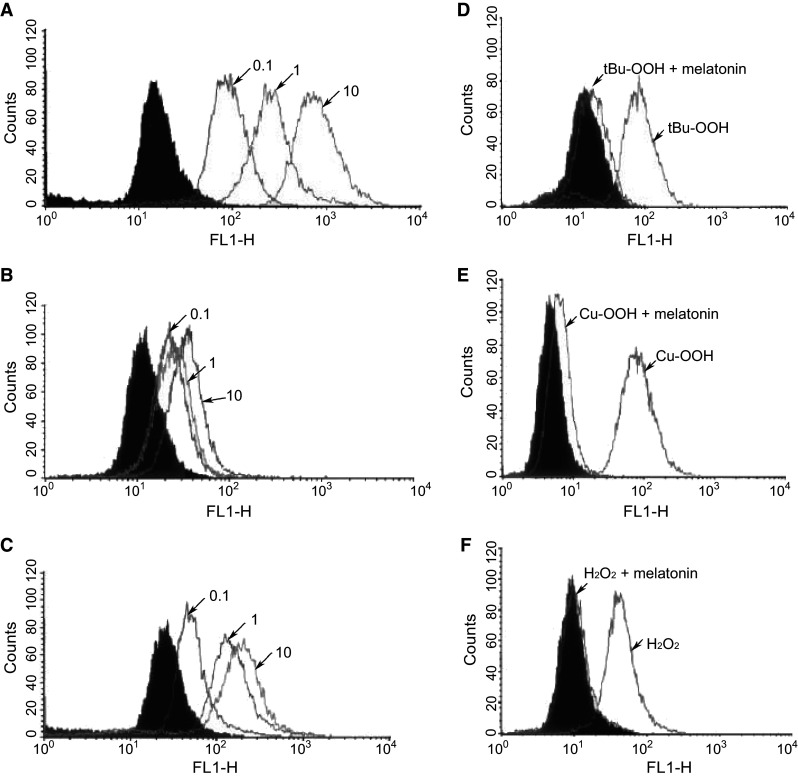
Fluorescence detected by flow cytometry (FACS) analysis of ROS generation in astrocytes after exposure to one of several oxidants: H2O2, tert-butyl hydroperoxide (t-BuOOH) or cumene hydroperoxide (Cu-OOH). a Illustrates the dose-dependent increase in mitochondrial ROS induced by H2O2 (0.1, 1.0 or 10 mM). b The addition of melatonin (100 µM) with H2O2 greatly diminished ROS fluorescence. c When 100 µM vitamin E was exchanged for melatonin, it was much less effective than melatonin in reducing mitochondrial ROS. d, e Melatonin also significantly lowered ROS generation in cells exposed to either t-BuOOH or Cu-OOH, respectively. f Melatonin inhibition of ROS-mediated by H2O2 exposure.
Reprinted with permission from Jou et al. [95]
