Fig. 3.
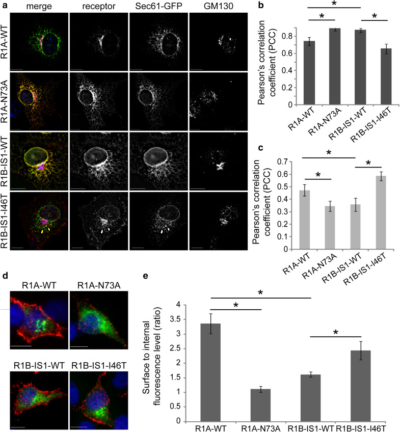
Glycosylation of Type-1 BMP receptors regulates their plasma membrane localization. a COS7 cells, co-transfected (24 h) with Sec61-GFP and myc-tagged Type-1 BMP receptors (R1A-WT, R1A-N73A RIB-IS1-WT or RIB-IS1-146T), were immunostained for myc-tag and GM130. Cells were imaged by fluorescence microscopy. Image stacks were de-convolved (NearestNeighbors deconvolution, SlidebookTM) and the PCC of different fluorescence channels was calculated. Micrographs depict a single focal plane of representative cells. Left column are merged images where receptors are in red, Sec6-GFP is in green and GM130 is in blue. Bar 10 µm. b, c Quantification of PCC of fluorescence signals of receptors and Sec61-GFP (b) or receptors and GM130 (c); n > 12 cells for each receptor construct. d Live COS7 cells transfected (24 h) with myc-tagged Type-1 BMP receptors (R1A-WT, RIA-N73A R1B-ISI-WT, or R1B-1S1-146T), were labeled in the cold with chicken-anti-myc and Alexa546-labeled donkey-anti-chicken antibodies, for detection of membrane-external receptors (red). Following fixation, samples were permeabilized and labeled with mouse-anti-myc and Alexa488-labeled goat-anti-mouse antibodies, for detection of internal receptors (green). Micrographs depict single confocal planes of representative cells. Bar 10 µm. e Quantification of the ratio of surface to internal fluorescence signals. Fluorescence signals were calculated from oonfocal z-stacks. Bar graph depicts averaged data (mean ± s.e.m.) of surface to internal ratio; n > 27 cells for each receptor construct

