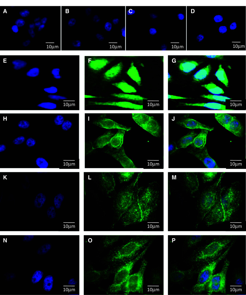
Fig. 3.

Cellular localization of TRPM7. Immunofluorescence was performed to localize TRPM7 in NT cells (a, e, f, g) and in cells expressing WT-CFTR (b, h, i, j), ΔF508-CFTR (c, k, l, m) and G551D-CFTR (d, n, o, p). First, negative controls in which first antibody is omitted shows the specificity of the labelling (a, b, c, d). Vesicular labelling in the cytoplasm is observed in all four cell types; however, membrane localization is mostly seen in cells expressing WT-CFTR (i, j) and ΔF508-CFTR (l, m). This membrane localization is likely not observed in NT cells (f, g). The labelling is less visible in ΔF508-CFTR (l, m) and lost in G551D-CFTR cells (o, p). Nuclei were DAPI stained (blue, a, b, c, d, e, h, i, j, k, l, m, n). Images were collected with a 100× objective. Bars represent 10 μm
