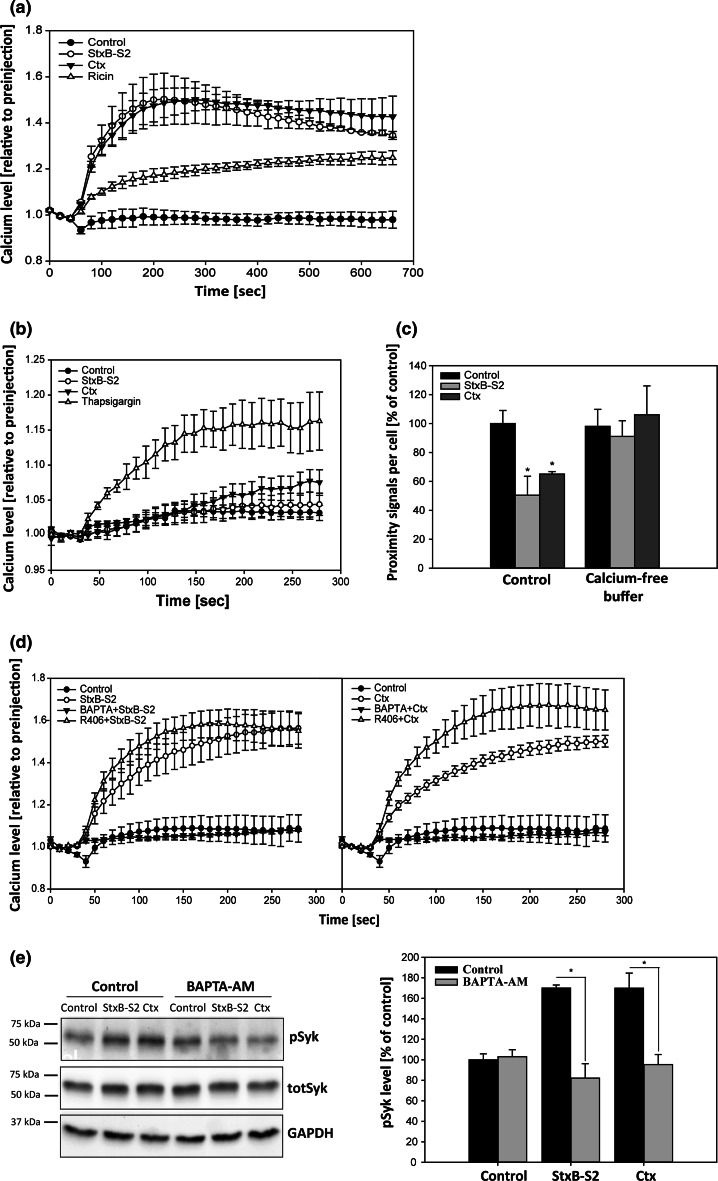
Fig. 4.
Shiga toxin and cholera toxin induce an increase in intracellular calcium, which is required for Syk activation. a HeLa cells were loaded with cell-permeable Fluo-4 for 30 min, followed by addition of 1 µg/ml ShigaB-sulf2 (StxB-S2), ricin or cholera toxin (Ctx). The fluorescence intensity was measured every 20 s for 10 min, and the data are normalized to the level of fluorescence prior to addition of the toxin. The mean values of at least three independent experiments are shown ±SEM. b The same experiment as in a, but performed in calcium-free buffer. In addition to StxB-S2 and Ctx, 1.5 µM thapsigargin (TG) was added. The fluorescence intensity was measured every 10 s for 4 min and the data are normalized to the level of fluorescence prior to addition of the toxin or TG (n = 3 independent experiments ±SEM). c HeLa cells were washed with Hepes-buffered medium (control) or calcium-free buffer before addition of 1 µg/ml StxB-S2 or Ctx, also in the indicated buffers, for 10 min. The cells were then fixed, permeabilized and subjected to PLA using cPLA2α and AnxA1 antibodies. Images were taken by an LSM780 confocal microscope, and the number of signals per cell was quantified and presented as % of control (n = 4 independent experiments, mean values +SEM, *p < 0.05). d Same as in a, but the cells were pre-treated with the indicated inhibitors for 30 min before addition of either 1 µg/ml StxB-S2 (left) or Ctx (right) and the fluorescence intensity was measured every 10 s for 4 min. The mean values of at least three independent experiments are shown ±SEM. e HeLa cells were transfected with a Syk-expressing plasmid for 24 h before being treated with 10 µM BAPTA-AM for 30 min followed by incubation with 1 µg/ml StxB-S2 or Ctx for 10 min. The protein lysates were subjected to immunoblot analysis using antibodies against phoshorylated Syk (pSyk), total Syk (totSyk) and GAPDH. Bar graphs show quantification of immunoblots with pSyk levels normalized to GAPDH and presented as % of control (n = 4 independent experiments, mean values +SEM, *p < 0.05)

