Fig. 6.
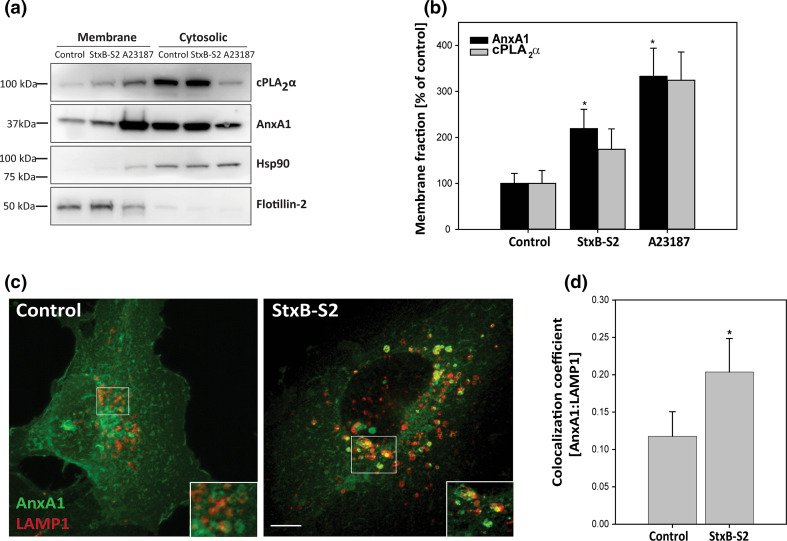
ShigaB-sulf2 induces translocation of AnxA1 to intracellular membranes. a HeLa cells were treated with 1 µg/ml ShigaB-sulf2 (StxB-S2) or 2 µM A23187 for 30 min, before the protein lysates were separated into cytosolic and membrane fractions and subjected to immunoblot analysis with antibodies as indicated. b Quantification of membrane fractions with AnxA1 and cPLA2α levels normalized to flotillin-2 and presented as % of control. The mean values of at least three independent experiments are shown as +SEM, *p < 0.05. c HMEC-1 cells were treated with 1 µg/ml StxB-S2 for 30 min before being fixed, permeabilized and subjected to immunofluorescence analysis using AnxA1 and LAMP1 antibodies. Images were acquired with an LSM780 confocal microsope, and the maximum intensity projection of 16 sections is shown. Scale bar, 5 µm. d Confocal images (single sections) obtained in experiments as described in c were analyzed, and the Manders colocalization coefficient between AnxA1 and LAMP1 was determined (n = 4 independent experiments, mean values +SEM, *p < 0.05)

