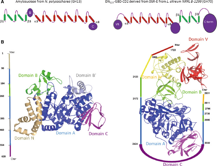
Fig. 2.
Structural characteristics of NpAS and ΔN123-GBD-CD2. a Topology diagrams of members of GH13 and GH70 families. Cylinders represents α-helices and β-sheets constituting the (β/α)8 barrel, and permuted elements are shown in green. b Schematic representation of the NpAS (PDB entry: 1G5A) and ΔN123-GBD-CD2 (PDB entry: 3TTQ) structures with labeling and color-coding of the five domains (A, B, and C domains common to GH13 and GH70 enzymes; N and B′ for amylosucrases, domains IV and V for GH70 members)

