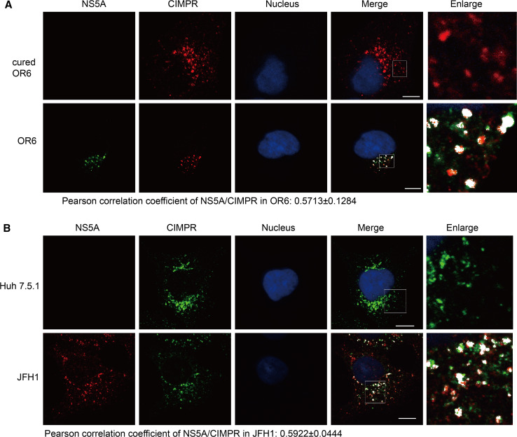
Fig. 6.
CIMPR localizes to the HCV replication area. Cured OR6, OR6 cells (a), Huh 7.5.1 cells or JFH1-infected Huh 7.5.1 cells (b) were fixed and immunofluorescently labeled for NS5A and CIMPR. DAPI marked nucleus. Areas of overlap are pseudocolored white in the merge and enlarge panels using an ImageJ colocalization highlighter plugin. The scale bars 10 μM

