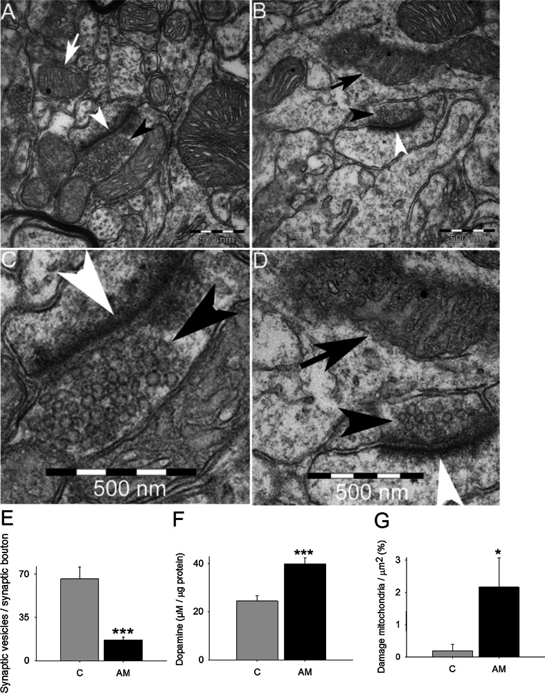
Fig. 4.
The effect of aminochrome on presynaptic monoaminergic vesicles in the neuron terminals. Aminochrome induces a significant decrease in the number of monoaminergic presynaptic vesicles in the terminals of dopaminergic neurons (b and d, a magnification b) in comparison with control animals (a and c, a magnification of a), determined using electron transmission microscopy in the striatum. The quantification of monoaminergic synaptic vesicles per terminal was plotted in e and the values correspond to the mean ± standard error. A significant increase in the amount dopamine was observed in isolated presynaptic vesicles of animals treated with aminochrome in comparison with control animals (f). In g is shown that aminochrome induced a significant increase in the number of damaged mitochondria per µm2 in animal treated with 1.6 nmol aminochrome (AM) in comparison to control animals in presynaptic terminals (c). The black head arrow shows synaptic vesicles. White head arrows show the synapses. White arrow shows normal mitochondria and black arrow shows damaged mitochondria. The inset is a magnification of synapses and synaptic vesicles in animals treated with aminochrome. The significance was measured with unpaired Student’s t test (***P < 0.001; *P < 0.05)

