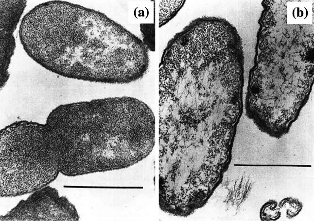
Fig. 11.

Transmission electron micrographs of a an untreated clinical isolate of Proteus mirabilis and b cells of the same isolate treated with 0.78 µg/mL (0.5×MIC) ampicillin for 3 h. The number of ribosomes has decreased following treatment. Bar 1 µm.
Images from [202] by permission of Proceedings of the Society for Experimental Biology and Medicine (SAGE Publications)
