
Fig. 2.
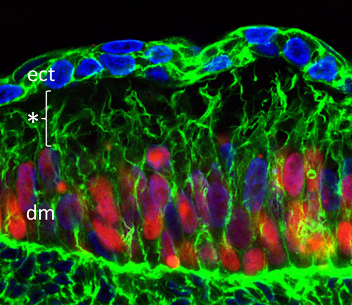
Signaling filopodia spanning the subectodermal space in the chicken embryo. Somitic cells (red) were labeled by overexpression of pCAGGS-H2B-mCherry-pA, nuclei (blue) are stained with DAPI, actin (green) was stained using Alexa Fluor 488 phalloidin. Filopodia are spanning the distance of about 20 µm (*) between the dermomyotome (dm) and the ectoderm (ect)