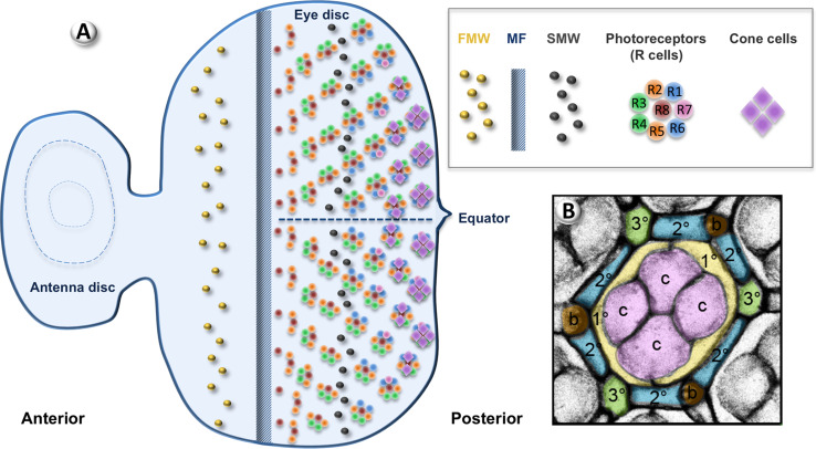
Fig. 2.
Eye development in Drosophila. a Schematic representation of a third instar larval eye-antenna disc. Anterior is to the left. Undifferentiated cells proliferate during the first mitotic wave (FMW). The morphogenetic furrow (MF) sweeps across the disc from posterior to anterior. Differentiation starts posterior to the MF with the recruitment of R8. Photoreceptors and cone cells are recruited sequentially. Ommatidia rotate on each side of the equator. The last round of proliferation occurs during the second mitotic wave (SMW). b From Martin-Bermudo et al. Apical view of a pupal retina 50 h after puparium formation stained with anti-Disc large (Dlg). The Dlg signal has been inverted so the staining appears in grey. At this stage, extra interommatidial cells have already been eliminated by apoptosis. The different cell types are pseudo-coloured for easier identification: cone cells (CCs) in pink, primary pigment cells (1°s) in yellow, secondary (2°s) and tertiary (3°s) pigment cells in blue and green, respectively, bristles in brown

