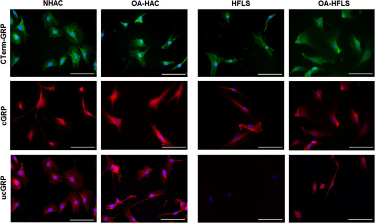
Fig. 2.
Accumulation patterns of GRP protein forms in both control (NHAC and HFLS) and OA (OA-HAC and OA-HFLS)-derived chondrocytes and synoviocytes. Immunofluorescence imaging was obtained for total GRP (CTerm-GRP), γ-carboxylated (cGRP) and undercarboxylated GRP (ucGRP) protein forms using specific antibodies. Cell nuclei were stained with DAPI; scale bar represents 100 µm. All experiments were repeated at least twice

