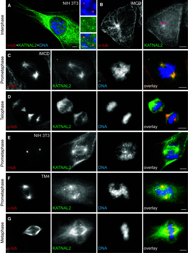
Fig. 3.

Intracellular localization of KATNAL2 on MTs, centrioles and mitotic spindle in different mouse cells by immunofluorescence. a, b At interphase, immunostaining for KATNAL2 (green) revealed a pronounced, detergent-resistant, cytoplasmic network and some concentration at the centrioles of the centrosome, as shown in NIH 3T3 fibroblasts (a; double labeling of centrosomes with anti-γ-tub in red; detail of centrioles at higher magnification in small side images) or IMCD cells (b; double labeling of MTs with anti-α-tub in red; arrowheads point at the two centrioles). Nuclei were counterstained for DNA with Hoechst (blue). c–f Series of examples of localization of KATNAL2 (green) during mitotic subphases, showing double labeling for α-tub (red) or for γ-tub (red, e) and counterstaining for DNA (blue) in different mouse cell lines (IMCD in c, d; NIH 3T3 in E; TM4 in f, g). KATNAL2 is highly concentrated at the centrosomes and MT asters at early mitotic phases and extends to the whole spindle in metaphase. The nucleocytoplasm is also strongly labeled throughout mitosis. Scale bars 5, 3 μm in detail of a
