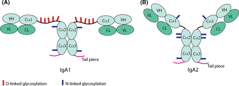
Fig. 3.
Schematic representation of the structural differences between human IgA1 and IgA2. The typical ‘T’ and ‘Y’ shape structure of IgA1 (a) and IgA2 (b), due to varying hinge length, are denoted in the figure, as are the light chain and heavy chain domains, namely, variable light (VL), constant light (CL), variable heavy (VH), the three heavy chain constant domains (Cα1, Cα2, Cα3) and the tail piece, which enables IgA polymerization. The difference in extent of and the positions of O-linked and N-linked glycosylation sites are shown with red and blue bars, respectively

