Fig. 11.
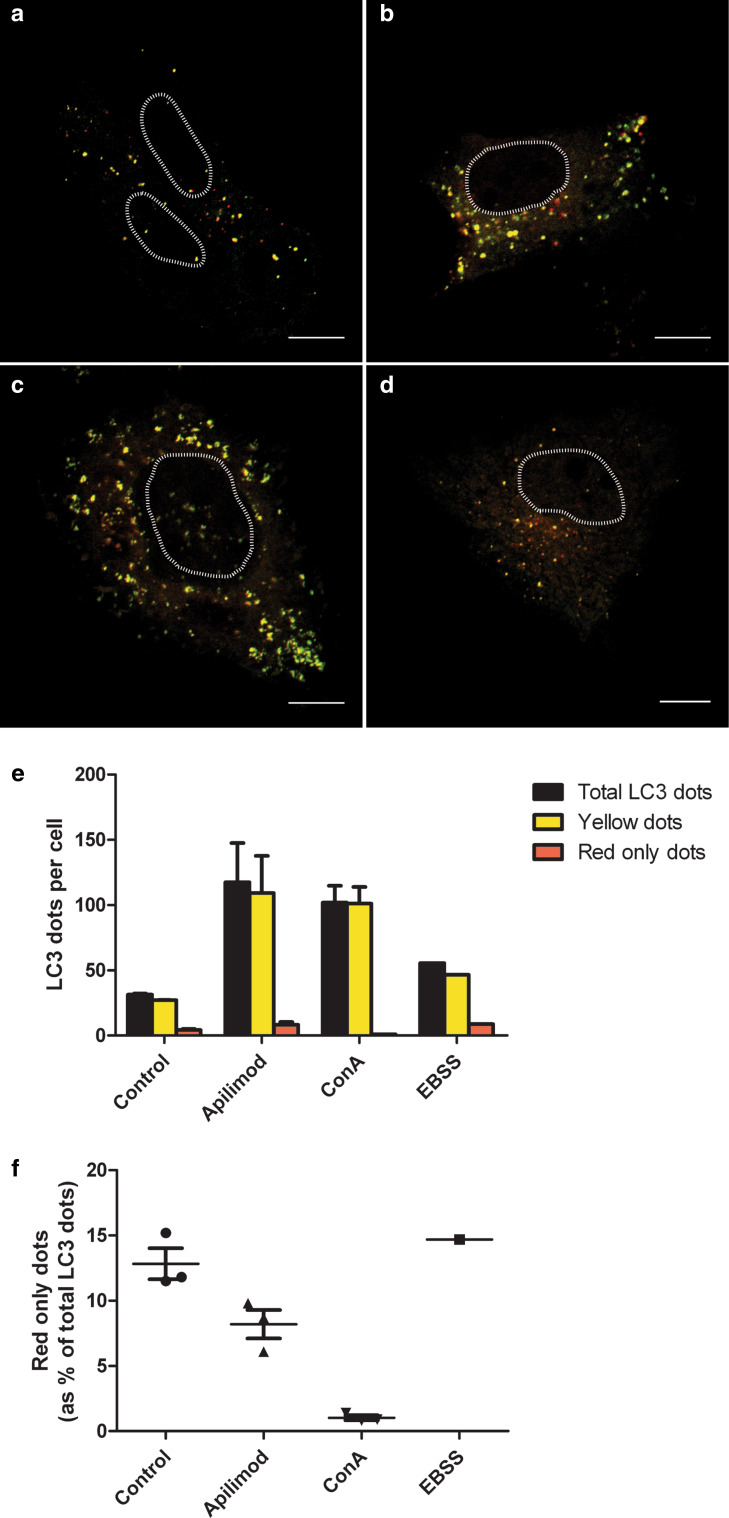
PIKfyve inhibition partly inhibits the fusion of autophagosomes with lysosomes. PC-3 cells were transfected with a double-tagged LC3 protein (mCherry-GFP-LC3), which will emit yellow (green merged with red) fluorescence in non-acidic structures and appear as red only in the autolysosomes due to quenching of GFP in these acidic structures. a Control cells, b cells treated with apilimod (0.5 µM) for 21 h, c cells treated with concanamycin A (50 nM) for 4 h and d cells grown in EBSS for 4 h. Nuclei are marked with a dashed line. Scale bar is 10 µM. e The number of yellow LC3 dots and red only LC3 dots per cell for each condition was quantified. Total LC3 dots are the sum of yellow and red only LC3 dots. f Percentage of red only LC3 dots of the total LC3 dots. From each experiment 12–22 cells were counted per condition and data are presented as mean values + standard error of the mean from 3 independent experiments. EBSS was included as a control, but only in one experiment

