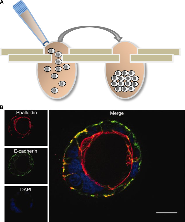
Fig. 3.
Three-dimensional (3D) techniques to culture colon cells. a 3D sphere formation using the hanging-drop technique. A cell suspension is applied to a specially designed plate (left) and cells accumulate and aggregate at the bottom of the drop. b 3D cyst formation by Caco-2 cells grown in Matrigel [101]. The confocal microscopy image shows DAPI-stained nuclei in blue and phalloidin-stained actin filaments in red, revealing the polarized cell organization and formation of a central apical lumen

